
MRI, CT, PET, and Ultrasound: What’s the Difference for Tumor Imaging?
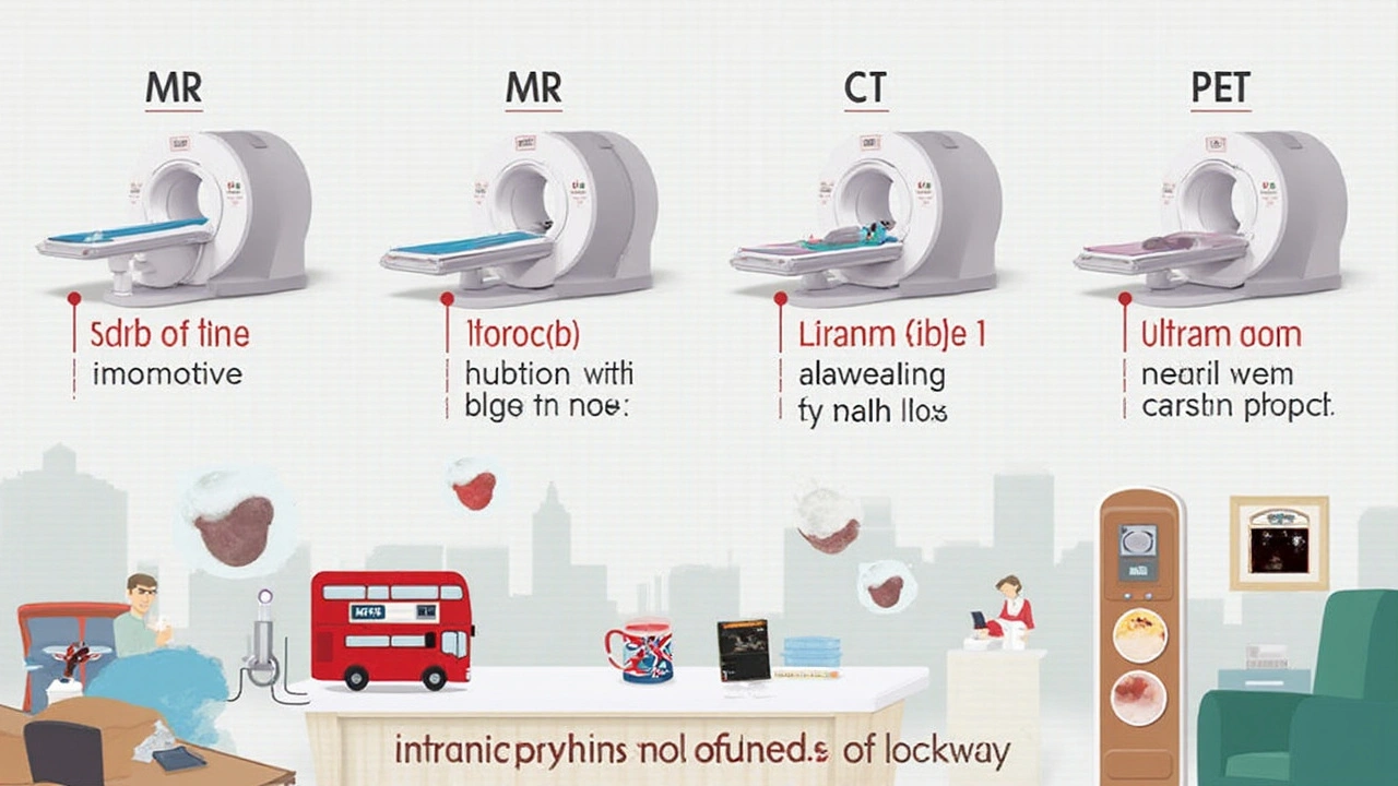
People throw around “MRI” and “CT scan” like they’re interchangeable, but when it comes to tumor imaging, the differences matter—big time. MRI (magnetic resonance imaging) uses powerful magnets and radio waves. It’s the go-to for soft tissues—think brain, spinal cord, liver—where you want every subtle detail. And if you’re watching for minute changes in a tumor’s outline, MRI’s resolution is tough to beat. That’s why it’s often used to check for regrowth in brain cancers or to keep an eye on how breast tumors shrink with chemo. There’s no radiation, so doctors can schedule repeat scans without stacking up a patient’s dose. But MRI is finicky. Metal implants can mess things up, and it’s a long, noisy, cramped process—sometimes not ideal for folks with claustrophobia, little kids, or anyone in serious pain who can’t keep still for thirty minutes straight.
CT (computed tomography) scans, on the other hand, are the workhorses in CT scan tumor size monitoring. They use x-rays to build a quick snapshot of body parts. You finish a CT in minutes, which is a lifesaver if a patient’s unstable or has trouble holding still. The catch: there’s radiation in every scan, and if you need regular tracking, that exposure adds up. Still, for lung, bone, and abdominal tumors, CT beats MRI in speed and can spot tiny calcifications MRI might miss. Most ER cancer checks or general follow-ups still rely on CT’s speed and broad coverage, even if the image isn’t as crisp for soft tissues.
PET (positron emission tomography) breaks the mold—it’s less about shape, more about what the tumor’s actually doing. You’re injected with a radioactive sugar tracer, and any rapidly-growing tumor (which eats sugar faster than healthy cells) will light up. This is powerful for knowing if odd-looking spots on other scans are just scar tissue or active cancer. Combining PET with a CT (called PET/CT) gives you the best of both—activity and structure. For some tricky situations, like lymphoma or metastatic disease, PET/CT can totally change the game.
Ultrasound is the simplest and most portable. You get instant feedback with no radiation. Ultrasound is king for certain types of tumors—like thyroid nodules, testicular lumps, or looking for changes in breast cysts. It’s totally safe for repeat checks and easy to use in an outpatient office—not just a hospital scanner room. Ultrasound’s main limitation is depth and operator skill; it doesn’t see through air or bone and heavily depends on who’s holding the probe.
Each tool brings its own set of strengths and quirks. Not every doctor—or patient—gets to choose the method for every scan. But knowing how they stack up helps you ask the right questions if you’re monitoring a tumor over weeks, months, or even years.
Which Imaging Is Most Accurate for Tumor Growth Monitoring?
If you want precision measuring—it’s not just about seeing the tumor, but also about charting how much it’s grown or shrunk. MRI tumor monitoring has a reputation for razor-edge accuracy, especially in the brain, spine, pelvis, liver, and soft tissues. MRI makers say you can spot changes as small as one to two millimeters. But accuracy isn’t guaranteed by the technology alone; it depends on clear scan protocols and consistent technique each time. For example, if a patient twists a little or breathes differently with each scan, the results can look way off. Some radiologists use software overlays to compare snapshots across time, but even then, a bad slice angle or fuzzy baseline image can skew measurements.
CT scan tumor size tracking is usually pretty solid for chest, abdomen, pelvis, and bones. One reason: CT’s slice-by-slice approach makes it simple to stack images and calculate three-dimensional measurements. Tumor boards (groups of doctors who review scans together) often rely on CT for deciding how a cancer responds to treatment, especially for lung or bowel cancers. One twist: tumors aren’t just round blobs, and CT doesn’t always capture shape changes as clearly as MRI.
PET scans don’t always deliver razor-sharp outlines. Their key strength is in tracking tumor activity—it’s the metabolic action, not just the mass, getting monitored. That means you might notice PET showing a tumor “shrinking” in terms of glucose uptake, even before it visually gets smaller. Many modern oncologists combine PET/CT for insight into both activity and bulk. In fast-moving cancers, PET can show whether a “shrunk” tumor is just scar tissue, or if there’s still active disease needing more therapy.
Ultrasound tends to lag behind when millimeter precision is needed for deep or challenging spots. That said, for certain accessible tumors—like liver, thyroid, or testicles—it can come close to CT or MRI, especially if the same tech does every check-up and uses digital calipers for the exact same measurement points.
Here’s a quick snapshot comparing key features:
| Imaging Modality | Accuracy | Radiation | Best For | Downsides |
|---|---|---|---|---|
| MRI | High, especially in soft tissue | No | Brain, spine, pelvis, liver | Long, noisy, expensive |
| CT | High, especially in chest/abdomen/bone | Yes | Lung, liver, bowel, bone | Radiation exposure |
| PET (PET/CT) | Moderate for size, excellent for activity | Yes | Lymphoma, metastatic cancer | Expensive, lower resolution |
| Ultrasound | Variable—excellent for superficial tissues | No | Thyroid, breast, testicle, liver (guided) | Operator-dependent, limited for deep/air-filled areas |
If you’re regularly tracking a tumor, stick to one imaging type whenever possible—switching back and forth muddies the data. A good radiologist will always mention if a scan’s quality limits the comparison, especially if tumor shape or location has changed. And keep an eye on low-level differences: some insurance plans or cancer centers favor one technology out of habit or budget, not because it’s actually best for you.
Radiation, Risks, and Patient Experience: What Matters for Long-Term Monitoring?
Radiation is where things get sticky. CT and PET both deliver ionizing radiation. For a single scan, the risk is tiny compared to the threat from untreated cancer. But lots of scans—sometimes monthly or even more often in tricky cancers—stack up. A chest CT can run you the equivalent of 100-300 standard x-rays, and PET/CT goes even higher. If you’re tracking a child’s tumor, anyone of childbearing age, or someone nearing full recovery, minimizing radiation exposure really matters.
That’s one reason MRI tumor monitoring and ultrasound hold such appeal—absolutely zero radiation. Children’s hospitals lean on MRI for brain or body tumors as much as possible for this very reason. Plus, the ability to rescan as often as needed is a massive win for high-stakes follow-ups.
Patient comfort is another huge deal and often gets overlooked by people who’ve never had to get scanned every month. MRIs take longer, require you to lie completely still in a tight tube (sometimes with a contrast agent injected), and can be so loud you need earplugs. CTs are quicker but still need an IV for contrast sometimes. PET requires you to get injected with radioactive sugar, sit perfectly still for an hour, then go through the scanner. With ultrasound, you might only need a simple gel squirted on your skin.
Safety for long-term monitoring isn’t just about radiation or noise. MRI is generally very safe—unless you’ve got metal in your body (like some aneurysm clips, pacemakers, cochlear implants), which can rule it out. Contrast used for MRI and CT can also cause reactions—rare, but something to think about if you have allergies, kidney issues, or previous reactions.
Children especially benefit from MRI or ultrasound, simply because the fewer the risks, the better. Adults often juggle multiple health issues, so sometimes CT’s speed or PET’s information outweighs the checklist of risks. It always comes back to what’s safest and most helpful for the person in the scanner—not just what the machine can do.
Best Practices, Recent Research, and Future Trends in Tumor Size Monitoring
Imaging has come a long way since blurry x-rays and guesswork measurements by ruler. Now, software can overlay images with millimetric precision, color-code tumor shrinkage, and even predict recurrence risk. Automated AI tools already help some radiologist teams flag growth that the human eye might miss, though they’re not perfect and require careful double-checking.
A highly cited study in 2022 compared MRI and CT tracking for head and neck tumors. It concluded that MRI beat CT for picking up early changes in soft tissue, but CT was more reliable for tracking overall size in bone or air-filled spaces. The best results? Combining sequences over several scans, always with the same technique.
If you or a loved one faces frequent scans, request access to the actual images (many hospitals have online portals now). That way, you and your healthcare team can spot trends or odd changes, not just trust summaries. Learn to keep a record of scan dates and findings. Sometimes hospital systems lose track of your imaging history, and having your own summary can prevent duplicated scans or mistaken conclusions.
Check out this detailed discussion on tumor growth monitoring to get a sense of how fast tumors can actually grow (hint: it varies a lot!). Understanding these differences can help you judge what kind of imaging intervals make sense.
One new advancement is the rise of diffusion-weighted MRI, which looks at water movement in tissue, giving early hints about a tumor’s response to therapy long before the mass actually shrinks. Ultrasonography with 3D and Doppler settings is getting smarter, letting doctors map out blood flow changes that show whether a tumor’s feeding vessels are shutting down or growing back. For rare tumors or complicated post-surgical cases, combining PET, MRI, and CT—called ‘hybrid imaging’—is sometimes the new gold standard.
Here are a few practical tips for patients facing regular imaging:
- Get familiar with your schedule—ask your team why they chose one scan over another.
- Let the scan tech know if you have metal, kidney problems, allergies, or claustrophobia. They can adjust technique or suggest alternatives.
- Try to use the same imaging center and radiologist for each scan where possible. Consistency matters.
- If you’re worried about radiation, ask if some scans can be replaced with MRI or ultrasound for interim monitoring.
- Always review the key findings with your oncologist. Not every finding means bad news, and not every shrinkage means cure. Context is everything.
In the future, expect more blends of imaging, digital overlays, and possibly even blood-based “liquid biopsies” to work alongside traditional scanning. For now, picking the right method—and using it well—makes all the difference for tumorous foes.


May 6, 2025 AT 21:57
Peter Aultman
Honestly, I've had three MRIs in the last year for a brain tumor. No radiation, but holy hell the noise. I brought headphones with classical music and still felt like my skull was vibrating. Worth it though. My doc says the detail saved my life.
Also, ultrasound for follow-ups on my liver? Surprisingly accurate when the tech knows what they're doing. Same person every time, same machine. That's the secret.
May 8, 2025 AT 15:51
Sean Hwang
CTs are fast but i hate the contrast shot. Feels like your whole body is on fire for a sec. My cousin got a PET/CT last month and they said the sugar thing made her dizzy. But man, it caught a spot the CT missed. So yeah, each tool got its moment.
May 9, 2025 AT 03:59
Barry Sanders
MRI is overrated. If your tumor isn't in a soft tissue zone, it's useless. CT is the real MVP. Stop pretending MRI is magic. And ultrasound? Please. That's for checking gallstones, not cancer progression.
May 9, 2025 AT 17:13
Chris Ashley
I just had my 8th CT in 14 months. My insurance denied an MRI because it's "too expensive." So now I'm basically a human radiation sponge. Why does this happen? Someone needs to fix this.
May 10, 2025 AT 02:06
kshitij pandey
In India, we don't always have access to MRI or PET. But ultrasound? We use it everywhere. My uncle's thyroid tumor was tracked for two years with just ultrasound and it worked fine. Same tech, same machine. Consistency beats fancy tech sometimes.
Also, no radiation = big win for families with kids.
May 10, 2025 AT 16:46
Brittany C
The literature is clear: longitudinal consistency in imaging protocol is paramount for accurate volumetric assessment. MRI with diffusion-weighted sequences demonstrates superior sensitivity for early treatment response in glioblastoma, whereas CT remains the standard for RECIST criteria in metastatic NSCLC. PET/CT is indicated for metabolic activity assessment, particularly in lymphoma.
May 11, 2025 AT 18:09
Sean Evans
People act like MRI is some kind of miracle. Nah. It's just expensive and slow. And don't get me started on patients who cry because it's "too loud." You think cancer is quiet?? 😤
CT is the real deal. Get over it. Also, if you're using ultrasound for anything beyond a thyroid nodule, you're doing it wrong. 🤦♂️
May 12, 2025 AT 00:51
Anjan Patel
I read this whole thing and I'm just mad. Why are we still using these 1980s technologies? Why isn't everyone getting a hybrid AI-enhanced quantum imaging scan? This is pathetic. You're all wasting time with CTs and ultrasounds when we could be using nanobots to map tumors in real time. The system is broken.
May 12, 2025 AT 15:27
Scarlett Walker
I'm a breast cancer survivor and MRI has been my best friend. No radiation, super detailed, and my oncologist says I'm lucky to have access to it. But honestly? The ultrasound tech who did my check-ups every 6 months? She was the real hero. Knew exactly where to press, how to hold the probe. That human touch? Priceless.
May 13, 2025 AT 05:25
Hrudananda Rath
It is, with profound regret, incumbent upon the modern medical establishment to acknowledge the abysmal inadequacy of conventional imaging modalities in the face of the escalating oncological crisis. The reliance upon antiquated, ionizing, and operator-dependent technologies constitutes a dereliction of scientific duty. One must question the epistemological foundations of radiological practice in the 21st century.
May 13, 2025 AT 12:21
Brian Bell
My dad's lung tumor was tracked with CT for 2 years. Then they switched to MRI and the doc said it looked like it shrunk 30%... but the CT before said it was stable. Turns out the MRI just had better contrast. So yeah, don't switch machines. Same center. Same tech. Same protocol. That's the real cheat code. 🤘
May 13, 2025 AT 13:15
Peter Aultman
^^^ This. I had a scan at two different hospitals and the measurements were off by 8mm just because the slice thickness was different. It's not the machine. It's the protocol. I started keeping a folder with the scan settings printed out. My oncologist was impressed. Small things matter.